
Fig. 10.
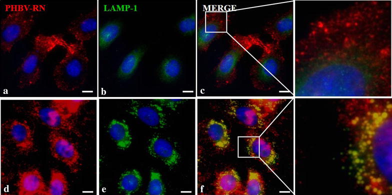
Determination of final nanoparticle fate in HeLa cells. HeLa cells were incubated with PHBV-RN nanoparticles for 15 min (a–c) and 1 h (d–f) at 37 °C; then cells were fixed and permeabilized. Later, an immunofluorescence was performed using anti-LAMP1 (1:100) and anti-mouse Alexa Fluor® 488 (1:500) as primary and secondary antibody respectively. Finally, cells were observed through fluorescence microscopy. Hoechst 33342 was used as nuclear stain. Objective: ×60 magnification. Bar size 10 µm