
Figure 8.
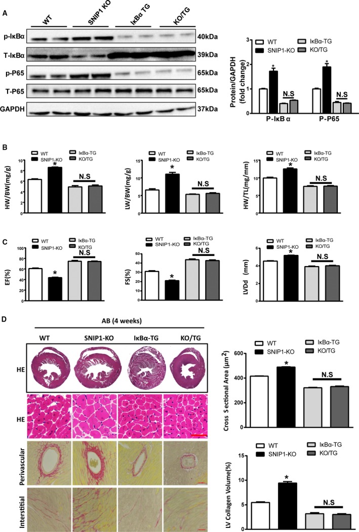
Overexpression of IκBαS32A/S36A ameliorates the pernicious effects of Smad nuclear interacting protein 1 (SNIP1) deficiency on aortic banding (AB)‐induced cardiac hypertrophy. A, Representative Western blot analysis and quantitative results of phosphorylated and total NF‐κB p65, inhibitor of NF‐κB α (IκBα) expression in wild‐type (WT), SNIP1‐knockout (SNIP1‐KO), IκBαS32A/S36A transgenic (IκBα‐TG), and SNIP1‐KO/IκBα‐TG (KO/TG) mice 4 weeks after AB operation (n=6 mice per group). B, Heart weight (HW)/body weight (BW), lung weight (LW)/BW, and HW/tibia length (TL) measured in the indicated mice 4 weeks after AB operation (n=10 mice per group). C, Results of ultrasound cardiogram test for the indicated mice at 4 weeks after AB operation, including fractional shortening (FS), ejection fraction (EF), and left ventricular end‐diastolic diameter (LVDd) (n=8 mice per group). D, Left: Hematoxylin–eosin (HE) staining and picrosirius red staining of the indicated mice hearts at 4 weeks after AB operation (n=5 mice per group; scale bar, 50 μm). Right: Bar graphs of calculated cross‐sectional area (n>100 cells per group) and left ventricular (LV) collagen volume (n>25 fields per group) in the indicated groups. Data are presented as the mean±SEM. *P<0.05 vs WT group; NS indicates no significant differences.