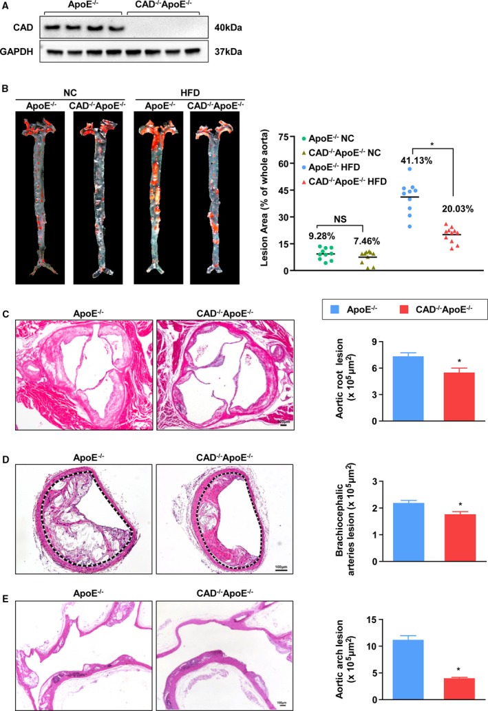
Figure 2.

CAD knockout alleviates development of atherosclerosis. A, Loss of CAD expression was confirmed by immunoblotting. B, Representative images of en face with Oil Red O staining of aortas of NC or HFD‐fed CAD −/−ApoE−/− and ApoE−/− littermates are shown in the left panel. Lesion occupation on the whole aorta was quantified (Right panel). n=10. *P<0.05 versus ApoE−/− HF group; NS, no significance. C through E, (Left) Representative images of hematoxylin and eosin–stained cross‐sections of aortic roots (n=10) (C), brachiocephalic arteries (n=4) (D), and aortic arches (n=3) (E). Scale bar=100 μm. (Right) Quantification of areas of atheromatous plagues. *P<0.05 versus ApoE−/− mice. CAD indicates caspase‐activated DNase; HF, high‐fat; HFD, high‐fat diet; NC, normal controls.
