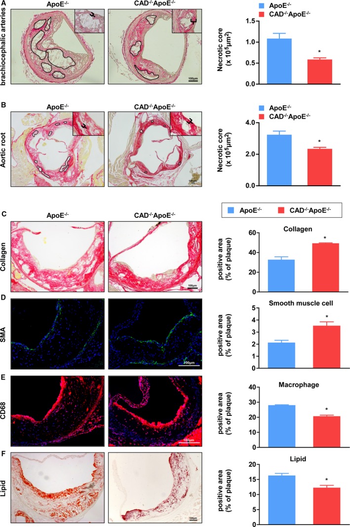
Figure 3.

Absence of CAD decreases necrotic areas and enhances stability of atheromatous plaques. A, (Left) PSR staining of histological sections of brachiocephalic arteries from CAD −/−ApoE−/− and ApoE−/− mice. (Right) Quantification of the areas of necrotic cores of brachiocephalic arteries (n=5). *P<0.05 versus ApoE−/− mice. B, (Left) Representative images of PSR staining of aortic roots from CAD −/−ApoE−/− and ApoE−/− mice. (Right) Quantification of the areas of necrotic cores of aortic roots (n=5). *P<0.05 versus ApoE−/− mice. C through F, (Left panel) Representative images of histological sections of aortic sinuses stained with PSR for collagen (C), SMA immunofluorescence for smooth muscle cells (D), CD68 for macrophages (E), and Oil Red O for lipids (F). Scale bar=100 μm. (Right panel) Atherosclerotic lesions from CAD −/−ApoE−/− mice displayed an increase in the percentage of collagen and smooth muscle cells, but a decrease in the percentage of macrophages and lipids compared to ApoE−/− mice (n=5). *P<0.05 versus ApoE−/− mice. CAD indicates caspase‐activated DNase; PSR, picrosirius red; SMA, smooth muscle actin.
