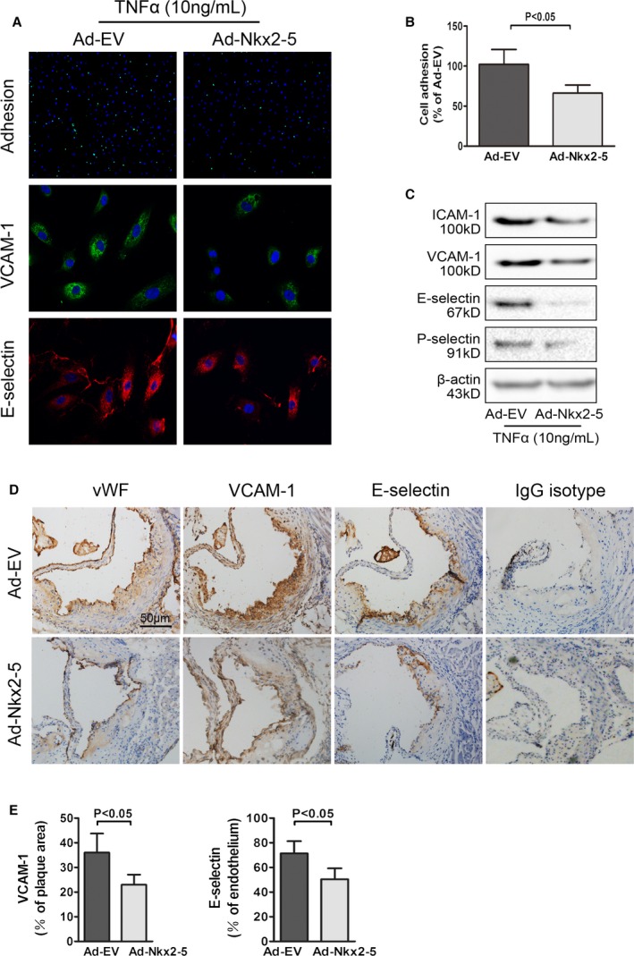
Figure 8.

NK2 homeobox 5 (Nkx2‐5) inhibits monocyte‐endothelial adhesion and decreases expression of adhesion molecules in early atherosclerosis. A, Upper panel: representative images used for quantification of peripheral blood monocytes (PBMC; green, calcein AM) attached to aortic endothelial cells (HAECs, blue, DAPI). Middle panel: effects of Nkx2‐5 on expression of vascular cell adhesion molecule‐1 (VCAM‐1) in endothelial cells (representative immunofluorescence image: green for VCAM‐1 and blue for cell nuclei stained with DAPI). Lower panel: effects of Nkx2‐5 on expression of E‐selectin in endothelial cells (representative immunofluorescence image: red for E‐selectin and blue for cell nuclei stained with DAPI). B, Quantification of adhesion. The number of PBMC adhered to per 100 HAECs was calculated. Results are expressed as a percent of values determined in the Ad‐EV‐treated group. Data represent the mean±SEM of 3 independent experiments. C, Representative immunoblot for intercellular adhesion molecule‐1 (ICAM‐1), VCAM‐1, E‐selectin, and P‐selectin in endothelial cells infected with Ad‐EV or Ad‐Nkx2‐5. D, Cross‐sections of early atherosclerotic lesions in aortic sinus were immunostained with antibodies against von Willebrand factor (vWF), VCAM‐1, and E‐selectin. Staining with rabbit IgG isotype was used as the negative control. E, Quantification of histochemical staining of VCAM‐1 and E‐selectin. Positive stained areas were quantified as a percentage of total plaque area or plaque endothelium. Data are expressed as mean±SEM (n=12 per group). AM indicates acetomethoxy; DAPI, 4′,6‐diamidino‐2‐phenylindole; HAECs, human aortic endothelial cells; IgG, immunoglobulin G; TNFα, tumor necrosis factor alpha.
