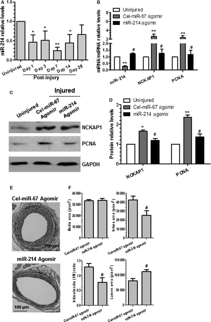
Figure 7.

Locally enforced expression of miR‐214 in the injured arteries inhibited NCK associated protein 1 (NCKAP1) expression levels, decreased smooth muscle cell proliferation, and blunted neointima hyperplasia after vascular injury. A, miR‐214 was downregulated after injury. *<0.05, **<0.01 (Compared with uninjured control). B through D, Gene/protein expression levels in the injured vessels were modulated by perivascular delivery of miR‐214 agomirs. After injury, 100 μL of 30% pluronic gel containing 2.5 nmol agomirs per vessel per mouse was immediately applied and packed around the injured vessel. At 3 days (B), 14 days (C and D), or 28 days (E and F) later, injured segments of femoral arteries were harvested and subjected to various studies, as indicated. Total RNAs and proteins were extracted from uninjured and injured vessels (femoral arteries from 3 to 5 mice were pooled for each experiment, n=3 experiments) and subjected to reverse transcriptase–quantitative PCR (B) and Western blotting (C and D) analyses. Representative images (C) and quantitative data (mean+SEM) (B and D) of 3 independent experiments are presented. E and F, Wire injury–induced neointima formation was blunted by miR‐214 overexpression. Paraffin sections from both groups (n=11 mice for each group) were prepared and subjected to hematoxylin and eosin staining analyses. Representative images (E) and quantitative morphological characteristics (mean+SEM) including media area, neointimal area, neointimal/media ratio, and lumen area (F) are presented. #<0.05 (Compared miR‐214 agomirs with Cel‐miR‐67 agomirs [B, D and F]). *<0.05, **<0.01 (Compared with uninjured vessels [B and D]).
