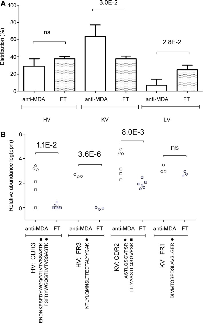
Figure 2.

Differences in the variable chain region between polyclonal anti‐MDA IgM and non‐anti‐MDA IgM (flow through, FT). A, Distribution in heavy variable (HV), kappa variable (KV), and lambda variable (LV) chains in the anti‐MDA and non‐anti‐MDA FT samples. B, Peptides from the HV and KV regions that were elevated in the anti‐MDA IgM. Numbers indicate significant P‐values. CDR indicates complementary determining region; FR, framework region; ns, not significant. MDA indicates malondialdehyde.
