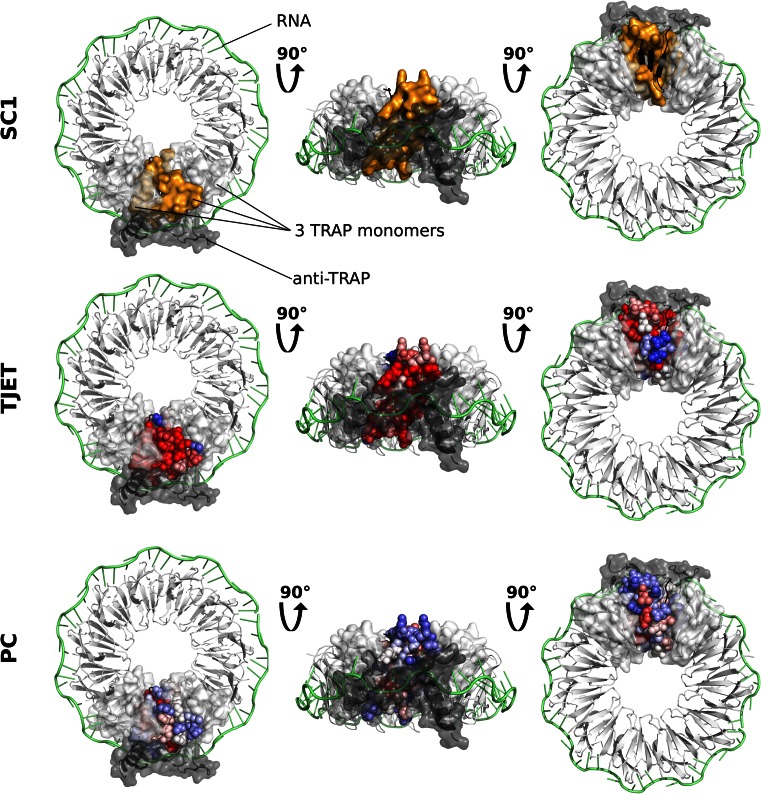
Figure 5.
JET2 predictions for the TRP RNA-binding attenuation protein (TRAP). Three different views (on the left, in the middle and on the right) of a homo 11-mer of TRAP in complex with a 53-base single-stranded RNA (1C9S) are shown. The RNA is colored in lime, 10 TRAP monomers are colored in white and 1 TRAP monomer is in black. The transparent surfaces of the two white monomers adjacent to the black one are displayed. A protein partner, anti-TRAP (2ZP8), is also shown as dark gray cartoon and transparent surface. The results of JET2 applied to TRAP (3ZZS) are mapped onto the structure of the black monomer: the binding site predicted by SC1 as orange surface, the values of the evolutionary trace TJET and of the interface propensities PC as blue through white to red spheres.

