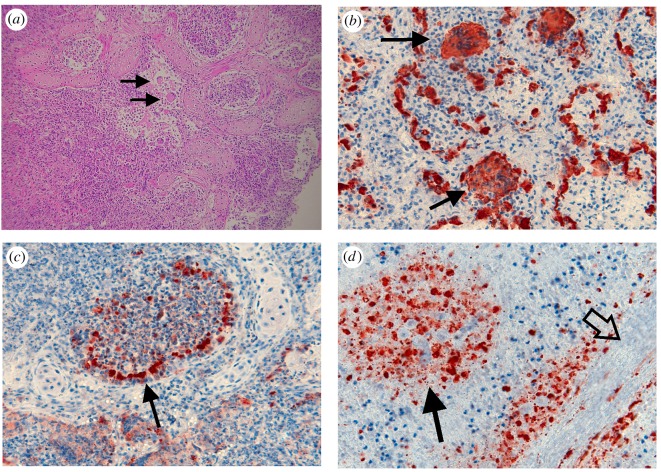
Figure 5.
Photomicrographs of lung and spleen sample from Indo-Pacific bottlenose dolphins during the 2013 UME. (a) Lung (H&E) showing extensive inflammatory cell infiltration. Bronchial and alveolar spaces filled with inflammatory cells, exfoliated epithelial cells and pneumocytes. Syncytia (solid arrows) present. (b) Lung (IHC for morbillivirus) showing immunostaining (brown) for viral antigen present in syncytia (solid arrows) and alveolar epithelium. (c) Lung (IHC for morbillivirus) showing immunostaining for viral antigen (brown) in bronchiolar epithelium (solid arrow), and in epithelial and inflammatory cells in bronchial lumen and lung parenchyma. (d) Spleen (IHC for morbillivirus) showing diffuse staining for viral antigen (brown) in white pulp (solid arrow) alongside splenic trabeculum (open arrow).

