Figure 2.
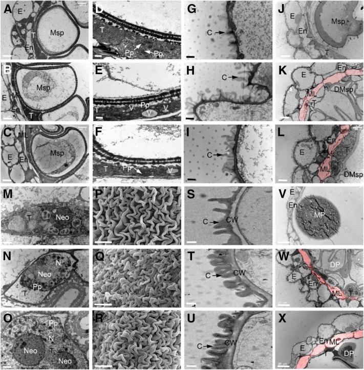
Electron microscopic examination of anther walls in wild type and dpw2 mutants. A to C, TEM images of anthers from the wild type (A), dpw2-1 (B), and dpw2-2 (C) at stage 10 showing the ring-vacuolate microspores. D to F, TEM images showing the morphology of tapetal cells of the wild type (D), dpw2-1 (E), and dpw2-2 (F) at stage 10. G to I, TEM images showing the outer region of anther epidermis in the wild type (G), dpw2-1 (H), and dpw2-2 (I) at stage 11. J to L, TEM images showing the anthers of the wild type (J), dpw2-1 (K), and dpw2-2 (L) at stage 11. M to O, TEM images showing the morphology of tapetal cells of the wild type (M), dpw2-1 (N), and dpw2-2 (O) at stage 11. P to R, SEM images showing the outer surface of anther at stage 12 in the wild type (P), dpw2-1 (Q), and dpw2-2 (R). S to U, TEM images showing the outer region of anther epidermis in the wild type (S), dpw2-1 (T), and dpw2-2 (U) at stage 12. V to X, TEM images showing the anthers of the wild type (V), dpw2-1 (W), and dpw2-2 (X) at stage 12. The cell layer highlighted in pink in K, L, W, and X represents the nondegraded middle layer in the mutant anther, which is absent in wild-type anthers at same stages. C, Cuticle; CW, cell wall; DMsp, degenerated microspore; DP, defective pollen; E, epidermis; En, endothecium; ML, middle layer; MP, mature pollen; Msp, microspores; N, nucleus; Neo, nucleolus; Pp, proplastid; T, tapetal layer; V, vacuole. Bars = 5 µm in A to C, J to L, and P to R; 1 µm in D to I, M to O, and S to U, and 10 µm in V to X.

