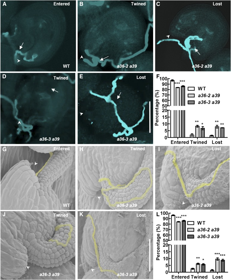
Figure 6.
Defective navigation of a36 a39 mutant pollen tubes in a limited pollination assay. A to E, Pollen tubes were stained with Aniline Blue and visualized by fluorescence microscopy. A, Wild-type (WT) pistils were pollinated with a limited number of pollen grains from the wild type. Wild-type pollen tubes enter the micropyle directly. B to E, Wild-type pistils were pollinated with a limited number of pollen grains from a36-2 a39 and a36-3 a39 plants. The pollen tubes were twisted around the micropyle (B and D), were growing on the ovule surface, or were twisted around the funicular surface, ultimately missing the micropyle (C and E). Bar = 50 μm. Arrows indicate the pollen tubes, and arrowheads indicate the micropyle. F, Statistical analysis of pollen tubes with different situations in limited pollination after 24 h by Aniline Blue staining. G to K, SEM analysis 24 h after pollination with a limited number of pollen grains. Pollen tubes are highlighted in yellow. Arrowheads indicate the micropyle. G, The normal pollen tube enters the micropyle after growth along the funiculus. H and J, The pollen tube was twisted around the micropyle. I and K, The pollen tube was growing on the integument, ultimately missing the micropyle. L, Statistical analysis of the different behaviors in pollen tubes with limited pollination after 24 h by SEM. For F and L, values are based on three biological replicates, error bars show se, and asterisks indicate values that differ significantly from those of the wild type (*, P < 0.05; **, P < 0.01; and ***, P < 0.001, calculated using two-way ANOVA).

