Figure 8.
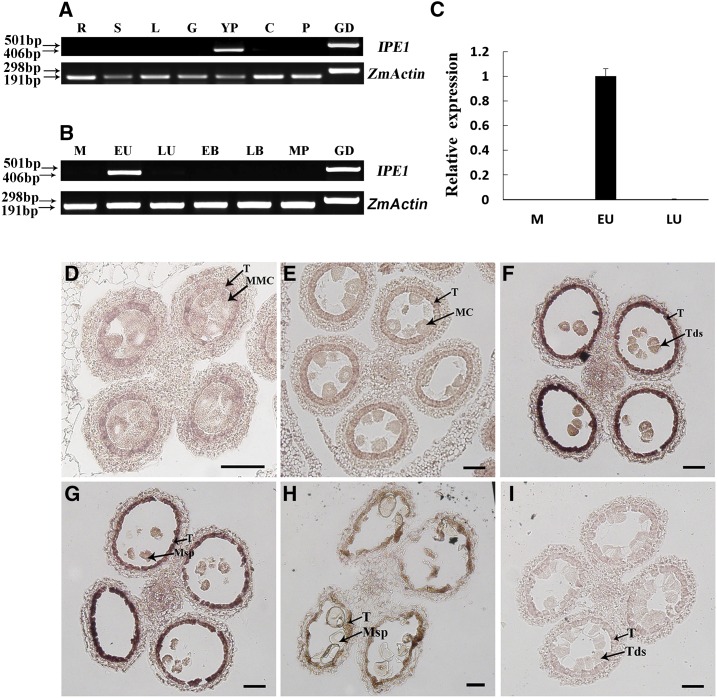
Expression analysis of the IPE1 gene. A, Detection of IPE1 transcript in wild-type selected tissues by RT-PCR, showing that IPE1 was expressed only in the young spikelets. ZmACTIN was used as a positive control. R, Roots; S, stems; L, leaves; G, glumes; YP, young spikelets; C, cobs; P, pistils; GD, genomic DNA. B, Detection of IPE1 transcript in wild-type anthers at different developmental stages by RT-PCR, showing that IPE1 was restricted temporally to the anthers during the early uninucleate microspore stage. M, Meiosis stage; EU, early uninucleate microspore stage; LU, late uninucleate microspore stage; EB, early binucleate microspore stage; LB, late binucleate microspore stage; MP, mature pollen grain stage. C, qRT-PCR analysis of IPE1 expression in wild-type anthers. Each data point is the average of three biological replicates. Error bars indicate sd (n = 3). D to I, RNA in situ hybridization of wild-type anthers using an IPE1-specific antisense probe (D–H) and a negative control sense probe (I). A strong hybridization signal in the tapetum was detected at the tetrad stage (F) and early uninucleate microspore stage (G). The signal in the tapetum disappeared at the early meiosis stage (D), meiosis stage (E), and late uninucleate microspore stage (H). Hybridization with the sense probe produced no signal at the tetrad stage (I). MC, Meiotic cell; MMC, meiotic mother cell; Msp, microspore; T, tapetum; Tds, tetrads. Bars = 50 μm.

