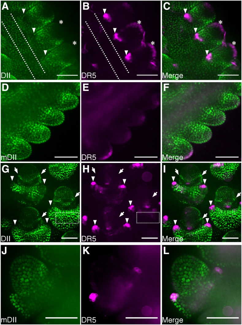
Figure 3.
DII and mDII signal in developing tassels. Fluorescence observed in secondary tassels excised from 5- to 6-week-old plants expressing DII (A and G) DR5 (B, E, H, and K), mDII (D and J), merged DII and DR5 (C and I), and merged mDII and DR5 (F and L). Imaged structures correspond to spikelet pair meristems (A–F) and floral meristems (G–I) and spikelet meristems (J–L). Bar = 100 μm. Asterisks indicate L1 layer. Arrowheads indicate glume primordia. Arrows indicate collar with high DII signal. Dotted lines in A and B and G and H indicate the central region of the tassel and the glume outside the spikelet meristem, respectively, that was used to quantify the fluorescence.

