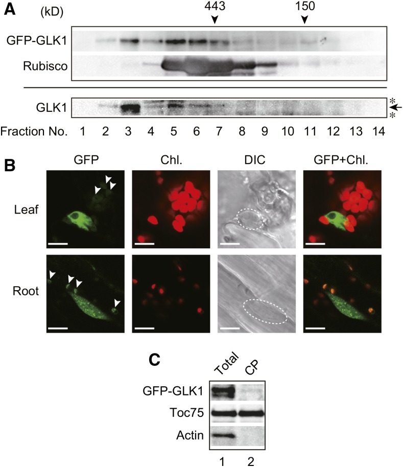
Figure 6.
Identification of GFP-GLK1 complexes by gel-filtration chromatography and intracellular localization of GFP-GLK1. A, Gel-filtration chromatography of GFP-GLK1 and GLK1 proteins expressed in Arabidopsis. Total protein extracts from GFP-GLK1 transformed glk1glk2 plants (upper panel) and GLK1ox plants (lower panel) were resolved by gel-filtration chromatography on a Sephacryl S-300 HR column. The molecular masses of the standard proteins are indicated by arrowheads. To verify the validity of chromatography, the distribution of Rubisco (approximately 550 kD) was also investigated. Proteins in each fraction were precipitated with trichloroacetic acid and analyzed by immunoblotting with antibodies against GLK1 or Rubisco holoenzyme. An arrow indicates the position of GLK1, and asterisks indicate nonspecific bands. B, Localization of GFP-GLK1 protein in Arabidopsis leaf and root cells. Leaf (upper) and root (lower) tissues of transgenic plants expressing GFP-GLK1 in glk1glk2 background were observed using a confocal laser-scanning microscope LSM 700. Dashed lines in DIC images indicate the location of the nucleus. Arrowheads indicate likely autofluorescence of plastids, rather than GFP signals. C, Detection of GFP-GLK1 in chloroplasts. Total protein extracts (lane 1) and chloroplast proteins (lane 2) were resolved by SDS-PAGE and probed with antibodies against GLK1 (top) and Toc75 (middle). As the negative control, the membrane was also probed with an anti-actin antibody (bottom). Chl., chlorophyll autofluorescence; DIC, differential interference contrast image; GFP, GFP fluorescence; GFP+Chl., overlap of the GFP and Chl. images.

