Fig. 5.
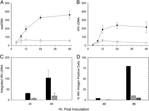
Replication of HIVNL4-3Δnef in CD4+ lymphoblastoid cell lines. The first round of replication for HIVNL4-3Δnef (○) in H9 cells was compared with HIVNL4-3 containing an intact nef gene (•) by real-time PCR to measure minus-strand strong-stop DNA synthesis (A) and completely synthesized HIV cDNA (B). (C) Integrated HIV cDNA (provirus) was also quantified for both HIVNL4-3Δnef (light-gray bars) and HIVNL4-3 (dark-gray bars). (D) Replication of two distinct clones of HIVNL4-3Δnef (light-gray and medium-gray bars) as measured by an immunofluorescence assay was also decreased compared with HIVNL4-3 (black bars) in MT-2 cells. In A–C, data are the average from three infections; error bars are one SD. Values are in equivalent numbers of chronically HIV-infected H9 cells (41, 42). Cells were infected with equal amounts of each clone based on input reverse-transcriptase activity.

