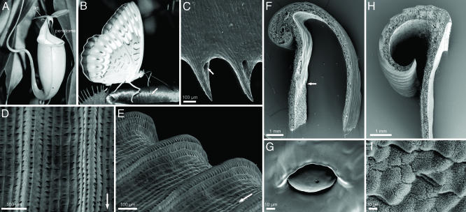
Fig. 1.
Nepenthes pitcher and peristome morphology. (A–G) N. bicalcarata. (A) Pitcher. (B) Butterfly (probably Tanaecia pelea pelea) harvesting nectar from the peristome surface. Note the visible line of peristome channels filled with nectar secreted from pores at the inner margin of the peristome (arrow). (C) Underside of inner margin of peristome with tooth-like projections and nectar pores (arrow). (D and E) Peristome surface with first- and second-order radial ridges. Arrows indicate direction toward the inside of the pitcher (F) Transverse section of peristome. Note the transition from the digestive zone to the smooth surface under the peristome (arrow). (G) Inner pitcher wall with digestive gland at the height of the inner peristome margin (H and I) N. alata.(H) Transverse section of peristome. (I) Waxy inner pitcher wall at the height of the inner peristome margin.

