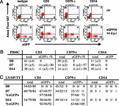
Fig. 3.
Time course and cell type specificity of CDV infections of PBMCs and lymphoid organs. (A) Fluorescence-activated cell sorter analysis of PBMCs isolated from an animal infected with 5804P-eGFP/H at 14 d.p.i. (Lower), and a noninfected control animal (Upper). Cells were stained with Alexa Fluor 647-labeled antibodies against cellular markers (y axis, see Mateials and Methods). eGFP expression was plotted on the x axis. (B) Percentages of CDV-infected T cells (CD3 positive), B cells (CD79α-positive), and MMs (CD14-positive). Groups of four animals were infected, and PBMCs were collected 0 (D0), 7 (D7), or 14 (D14) d.p.i. The mean values are indicated, with SD shown parenthetically. Total cells include eGFP-positive (eGFP+) cells. (C) CDV-infected cells in the lymph nodes (LN), spleen (SP), or thymus (TY) of animals killed 7 (D7) or 14 (D14) d.p.i., or a control animal (D0). In the lines labeled %eGFP+, the percentages of GFP-expressing (CDV-infected) cells within one cell type are indicated.

