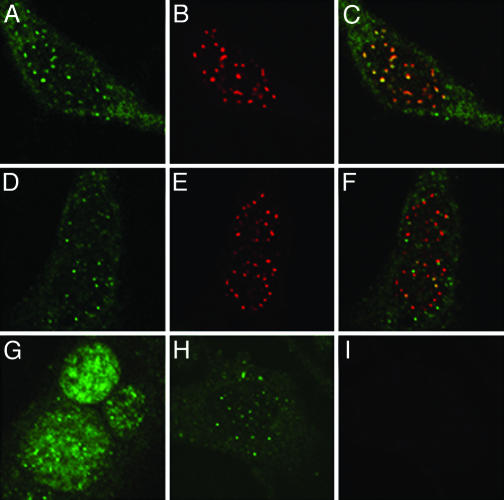
Fig. 5.
L2 and pseudogenome localization in HP3 and Hemp cells. Cells were allowed to internalize L1 + L2-HA pseudovirions that were assembled in the presence of 20 mM BrdUrd. Forty-eight hours postentry, the cells were fixed and processed for BrdUrd detection coincident with detection of the HA epitope and PML protein. (A–C) Staining of the PML3-expressing HP3 cells to show the distribution of the L2 protein, detected with an anti-HA monoclonal antibody, is shown in A; the rabbit anti-PML staining is shown in B; and the merge of the two channels is shown in C. D–F also show the HP3 cells. D, mouse anti-BrdUrd detection; E, anti-PML staining; and F, the merge. Staining of the pseudovirion components in Hemp cells is shown in G–I. G, the more diffuse localization of L2, represented by anti-HA detection; H, the pattern of the BrdUrd-labeled pseudogenome; I, the anti-PML staining of these cells demonstrating the lack of expression of this protein.

