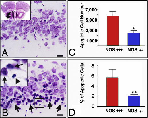
Fig. 4.
Confirmation of the significance of nNOS/NO signaling for transsynaptic apoptosis in piriform cortex with bulb lesions in nNOS–/– mice. (A Inset) A comment on the anatomy of the mouse olfactory bulb. (B Inset) A magnification of the framed area in B. (A and B) In WT mice, bulb lesions cause transsynaptic degeneration of layer IIα neurons 24 h after lesion. Compare a sham-operated (A) with a bulbectomized (B) animal. Degenerating neurons (B) show shrinkage and pyknosis (arrows in Inset); fragmentation of the nucleus is also evident in some cells (arrowhead in Inset). As shown in A Inset, a substantial portion of the mouse olfactory bulb is located underneath the frontal pole (arrows); this anatomical feature makes it necessary to employ careful aspiration lesions, as explained in Materials and Methods. (C and D) In NOS–/– mice, piriform neurons undergoing transsynaptic degeneration after bulbectomy are significantly reduced in both number (C) and percentage in the total number of layer I-II pyramidal neurons. (D). *, P = 0.0167; **, P = 0.0015. (Scale bars, 20 μm.)

