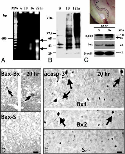
Fig. 6.
Evidence of free radical release, DNA injury, and cytosolic signaling of apoptosis on microdissected samples of layer II piriform cortex. (A Inset) The layer II-specific dissection, as compared to layer I dissection illustrated in Fig. 3C. (A) DNA blots show fragmentation in nucleosomal multimers. There are ≈200- and ≈400-bp multinucleosomal bands 22 h after bulbotomy (arrows), but some nonrandom fragmentation is also seen by 16 h. (B) Oxyblot detection of extensive carbonylation of proteins in layer II of piriform cortex 10–12 h after bulbotomy. (C) Twelve hours after bulbotomy, i.e., at a time point overlapping with oxidative injury as in B and upstream to DNA fragmentation (A), Western blotting shows evidence of DNA injury based on PARP cleavage and cytosolic signaling of apoptosis based on bax up-regulation. (Upper) The layer-II specific dissection that generated samples for analyses in A–C; compare with layer I dissection illustrated in Fig. 3C. (D) At 14–20 h after bulbotomy, bax protein accumulates in layer II neurons (arrows) at levels detectable by ICC. (Upper) Because of space limitations, the image was edited by removing the middle and approximating the medial and lateral portions of the section. (E) Twenty hours after bulbotomy, several layer IIα profiles developed strong immunoreactivity to active caspase 3 (arrows). (Upper) Two bulbotomy cases. (Lower) The sham condition. Specific immunoreactivity is localized to the cytoplasm. Some nonspecific nuclear staining is present in many cells both in the bulbotomy and sham scenarios. Bx, bulbotomy; S, sham. (Scale bar, 20 μm.)

