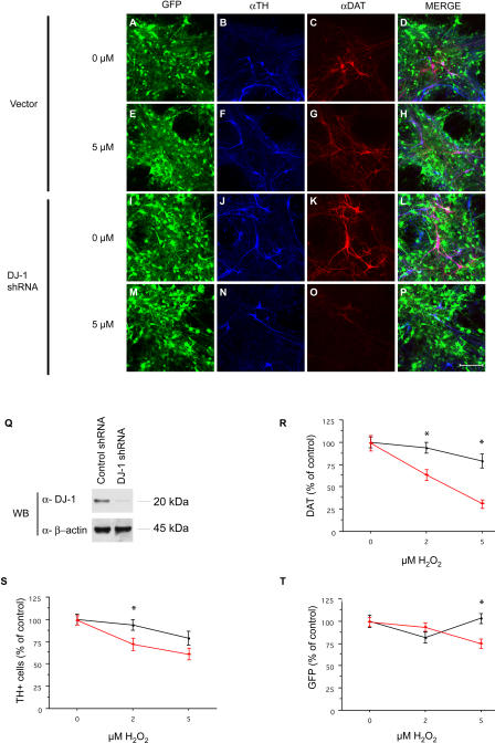
Figure 5. RNAi “Knockdown” of DJ-1 in Primary Embryonic Midbrain DNs Display Increased Sensitivity to Oxidative Stress.
(A–P) Primary midbrain cultures from E13.5 embryos were infected with lentiviral vectors encoding DJ-1 shRNA (or vector alone) under the regulation of the control vector (A–H) or the U6 promoter (I–P). Cells were cultured for 1 wk after infection and then exposed to H2O2 (5 μM; E–H and M–P) for 24 h. Cultures were immunostained for TH (B, F, J, and N) or DAT (C, G, K, or O) and visualized by confocal microscopy. Images containing all stains are included (Merge; D, H, L, and P). Scale bar, 100 μm.
(Q) Cell lysates prepared from midbrain primary cultures infected with DJ-1 shRNA lentivirus (or control vector) were analyzed by Western blotting for murine DJ-1 or β-actin.
(R–T) Quantification of TH, DAT, and GFP signal was performed on ten randomly selected fields in each of three wells for each condition. Red triangles, DJ-1 shRNA treated; black circles, control vector. Data represent the means ± SEM and were analyzed by ANOVA followed by Fisher's post-hoc test. * p ≤ 0.05.

