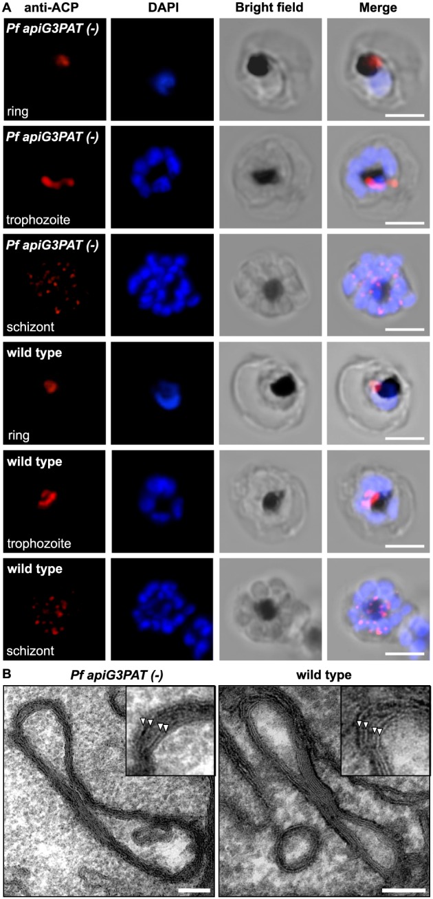
Figure 2.

Pf apiG3PAT (−) parasites have normal apicoplast morphology and development in the blood stage. A. Immunofluorescence microscopy of Pf apiG3PAT (−) and wild type parasites using antibodies against the apicoplast marker ACP demonstrates disruption of the enzyme has no effect apicoplast morphology in rings, trophozoites or schizonts. DNA stained with DAPI. Scale 3 µm. B. Transmission electron microscopy of trophozoite stage Pf apiG3PAT (−) and wild type parasites confirms disruption of the enzyme has no effect on apicoplast structure or membrane organization. Magnified images (inset) show the four apicoplast membranes are arranged in pairs (arrowheads) in both lines. Scale 100 nm.
