
Figure 4.
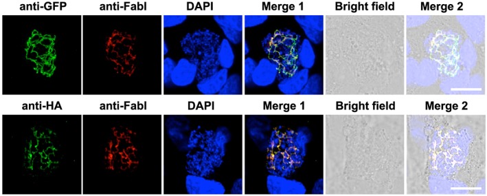
Pb apiG3PAT is expressed in liver stage parasites and localizes to the apicoplast. Immunofluorescence microscopy of Pb apiG3PAT ha gfp liver stage parasites 48 h post‐infection confirms the GFP and HA signals co‐localize with the apicoplast marker FabI. Parasite and host cell nuclei stained with DAPI. Scale 10 µm.