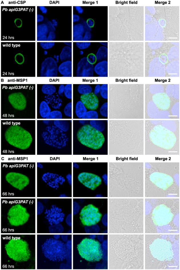
Figure 8.

Pb apiG3PAT (−) parasites show altered plasma membrane morphology in the late liver stage but still express merozoite surface protein 1. A. Immunofluorescence microscopy of Pb apiG3PAT (−) and wild type parasites using antibodies against the CSP at 24 h post‐infection. Pb apiG3PAT (−) and wild type parasites show similar patterns of CSP staining at this time point, indicating plasma membrane morphology is not affected by disruption of the enzyme in the early liver stage. B. Immunofluorescence microscopy of Pb apiG3PAT (−) and wild type parasites using antibodies against merozoite surface protein 1 (MSP1) at 48 h post‐infection. Similar patterns of MSP1 expression are observed in Pb apiG3PAT (−) and wild type parasites, indicating disruption of the enzyme does not markedly affect plasma membrane morphology or MSP1 expression at this stage. C. Immunofluorescence microscopy of Pb apiG3PAT (−) and wild type parasites using anti‐MSP1 antibodies at 66 h post‐infection. Pb apiG3PAT (−) and wild type parasites differ noticeably in their pattern of MSP1 staining at this time point, suggesting disruption of the enzyme affects formation of merozoite membranes in the late liver stage. Importantly, although there was only one instance where evidence of plasma membrane invagination was observed for Pb apiG3PAT (−) parasites (middle panel), MSP1 expression was still readily detected in the line. DNA with DAPI. Scale 10 µm.
