
Figure 9.
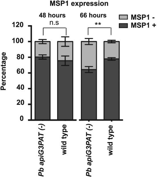
Pb apiG3PAT (−) parasites show only a minor defect in merozoite surface protein 1 expression in the late liver stage. Proportion of Pb apiG3PAT (−) and wild type parasites expressing merozoite surface protein 1 (MSP1) at 48 and 66 h post‐infection. Pb apiG3PAT (−) and wild type parasites do not differ significantly in the proportion of MSP1 positive parasites at 48 h post infection as determined by a two tailed t‐test (n.s., non‐significant). The proportion of MSP1 positive parasites in the Pb apiG3PAT (−) and wild type lines does differ significantly at 66 h post‐infection (**, p‐value <0.05), but as still over 60% of Pb apiG3PAT (−) parasites express MSP1 at this time point, this defect appears to be relatively minor. Error bars show mean of three biological replicates ± standard error.