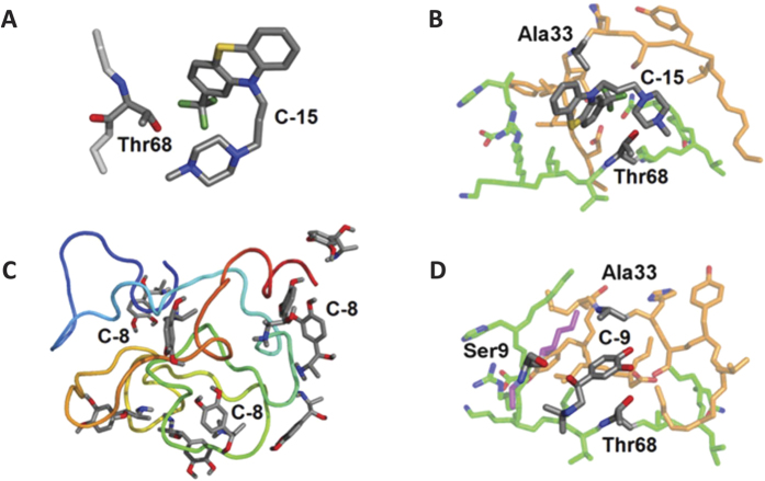
Figure 4. Docking of compounds to NUPR1.
(A) Binding mode of Compound-15 with the side chain of Thr68–a portion of the protein main-chain (light gray) is also shown. (B) A transient protein pocket modelled by combining binding modes of Compound-15 with NUPR1 main chain portions including residues 27–40 (orange) and 62–72 (green). (C) Numerous binding modes of Compound-8 with the protein main chain (N and C terminus in blue and red, respectively). (D) A transient protein pocket of Compound-9 with NUPR1 main chain portions including residues 8–11 (purple), 27–40 (orange) and 60–72 (green). PyMol57 was used for all displays; hydrogens and protein main-chain oxygens are not shown, and protein backbone nitrogens are colored only for labelled residues.

