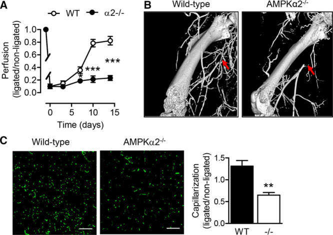
Figure 1.

Recovery of blood flow and revascularization in ischemic hindlimbs after femoral artery ligation in diabetic and AMPKα2−/− mice. A, Reperfusion after femoral artery ligation as measured by laser Doppler analysis in wild-type (WT) and AMPKα2−/− littermates (n=9 per group). B, Micro computed tomography angiography of ligated limbs from WT and AMPKα2−/− mice 7 d post surgery. Arrows indicate site of ligation. Similar results were obtained in an additional 2 animals in each group. C, CD31+ capillaries in transverse sections of ischemic gastrocnemius muscles from WT and AMPKα2−/− mice (bar=100 µm; n=4 per group). *P<0.05, **P<0.01, ***P<0.001 vs control group. AMPK indicates AMP-activated protein kinase.
