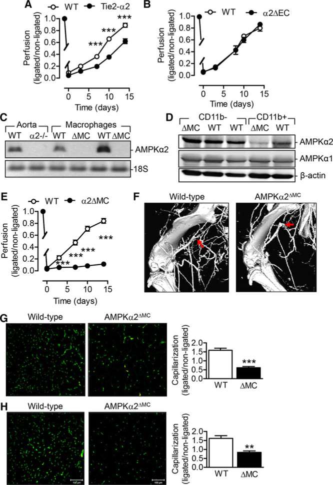
Figure 2.

Comparison of the consequences of endothelial cell and myeloid cell AMP-activated protein kinase (AMPK) α2 deletion on vascular repair. A and B, Recovery of blood flow after femoral artery ligation in hindlimbs from (A) wild-type (WT), Tie2-α2 littermates (n=8 per group), and (B) WT and AMPKα2ΔEC littermates (n=4 per group). C, AMPKα2 mRNA expression in aorta and macrophages isolated from WT, AMPKα2−/− or AMPKα2ΔMC mice. Similar results were obtained in 2 additional experiments. D, AMPKα2 and AMPKα1 protein expression in CD11b− and CD11b+ cells isolated from spleens from WT and AMPKα2ΔMC littermates. Similar results were obtained in 2 additional experiments. E, Recovery of blood flow after femoral artery ligation in hindlimbs from WT and AMPKα2ΔMC mice (n=9 per group). F, Micro computed tomography angiography of ligated limbs from WT and AMPKα2ΔMC mice, 7 d post surgery. Arrows indicate site of ligation. G and H, Anti-CD31 immuno-stained capillaries (green) in the semimembranosus (G) and gastrocnemius (H) muscles of ligated limbs from WT and AMPKα2ΔMC mice 14 d after surgery. The quantification was performed relative to the nonligated limb (n=5 per group). *P<0.05, **P<0.01, ***P<0.001 vs WT.
