
Figure 2.
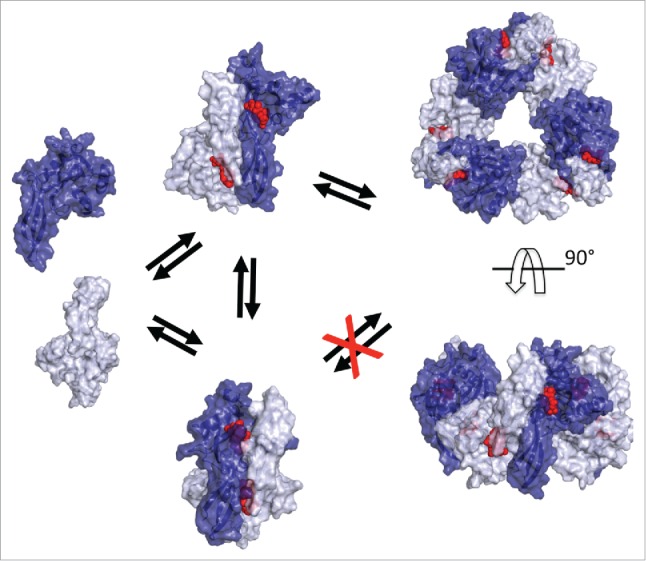
Model of NUDIX5 dimers, and hexamer. From left to right: 1) Surface representation of NUDIX5 monomers (light and dark blue); 2) Two versions of the NUDIX5 homodimer: the published one (PDB code 2DSC) at the bottom cannot bind PPi as required for making ATP; the “flipped” conformation at the top facilitated by T45 dephosphorylation could bind PP1; 3) in silico predicted NUDIX5 homo-hexameric structure based on the “flipped” homodimer, top view (top) and side view (bottom). The model of NUDIX5 hexamer was obtained using MODELLER and refined with ROSETTA. Structures shown in the figure were rendered using PyMOL (http://www.pymol.org). ADPR is shown in red.