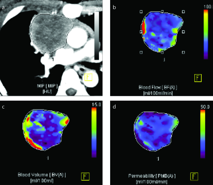
Figure 2.
a–d. A 44-year-old patient with a pathologic diagnosis of lymphoma. The MIP image (a) shows a heterogeneous off-midline mass at the anterior mediastinum. Corresponding blood flow map (b) shows heterogeneously decreased blood flow in the lesion. Blood volume map (c) also shows low values with an inhomogeneous distribution. Corresponding permeability surface map (d) shows areas with homogeneously decreased permeability within the lesion.

