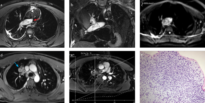
Figure 2.
a–f. A-30-year-old man with intimal sarcoma. On transversal (a) and coronal (b) fat-suppressed T2-weighted imaging, the dilated filling defect in the right pulmonary artery is hyperintense and extends to the right upper lobe pulmonary artery (red arrow). DWI (b=800 s/mm2) (c) shows the lesion as mildly hyperintense. Contrast images (d, e) show that the lesion filled the right upper lobe pulmonary artery along its course giving it a grape-like appearance (blue arrow); the lesion is significantly enhanced (e). Histopathology (f) demonstrates an abundance of malignant spindle cells with high cellularity and a high nuclear/cytoplasmic ratio (HE staining, ×200).

