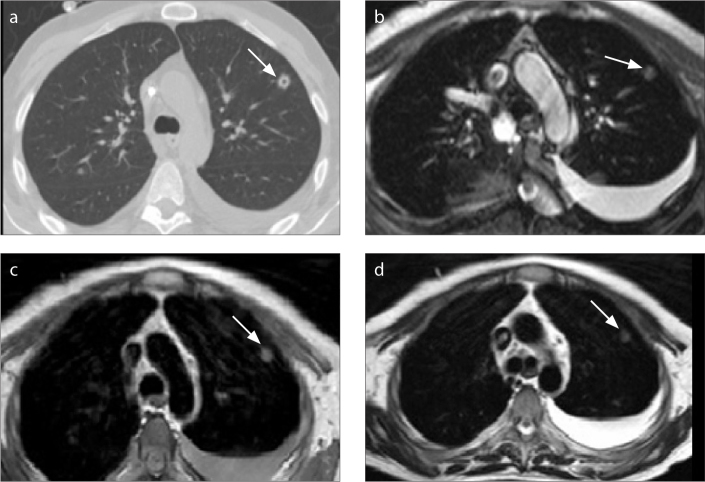
Figure 4.
a–d. A 38-year-old man with Burkitt lymphoma and invasive pulmonary aspergillosis. CT image (a) shows cavitating nodule (arrow) in the left upper lobe and left pleural effusion. B-FFE (b), T1-weighted (c), and T2-weighted (d) images show a nodule (arrow) and the pleural effusion. The cavity in the nodule is not seen on MRI.

