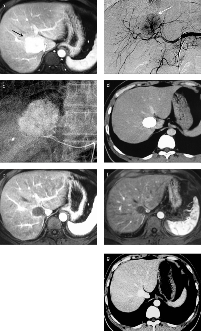
Figure 2.
a–g. A 34-year-old male patient suffering from symptomatic FNH of the liver. Preprocedural enhanced MRI in the arterial phase (a) demonstrates an enhancing mass (arrow) located in segment VIII. This mass increased in size compared with an earlier examination. Celiac arteriogram (b) demonstrates a hypervascular mass (arrow) that is supplied by a dominant feeding artery radiating in a spinning wheel pattern from the periphery into the mass. (c) The feeding artery was selectively catheterized and embolized with a microcatheter. CT imaging (d) performed at three-month follow-up demonstrates significant reduction in lesion size and good uptake of iodized oil in the lesion. Contrast-enhanced MRI exams performed at six-month (e) and 24-month (f) follow-ups show no residual enhancement of the mass, which has slightly decreased in size. Arterial phase CT image (g) obtained at 36-month follow-up demonstrates further slight decrease in size.

