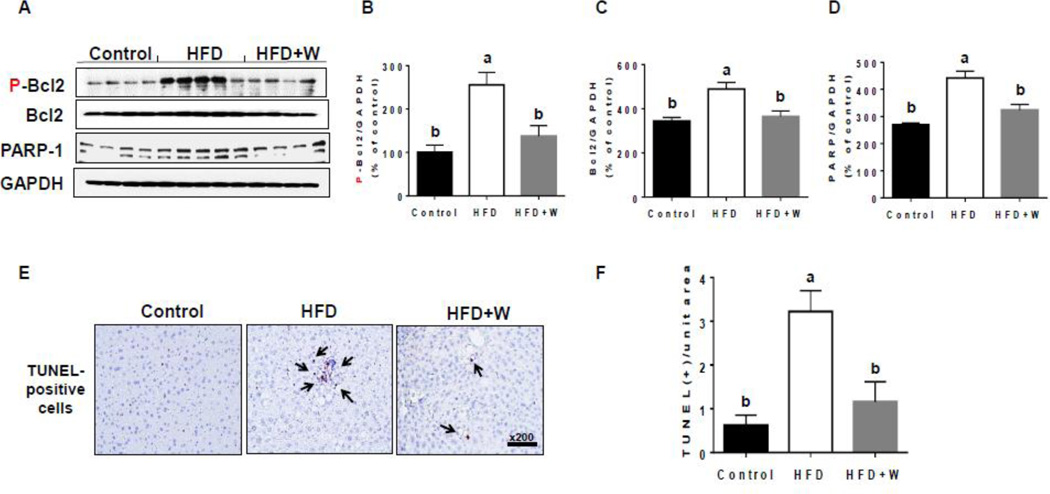
Figure 7.
Effects of dietary walnuts on the hepatic apoptosis. (A) Representative images of the immunoblot analysis for P-BCL-2, BCL2, PARP-1 or GAPDH in the livers of experimental mice exposed to HFD or HFD+walnut for 6 weeks. The densitometric levels of (B) P-BCL2, (C) BCL2, and (D) PARP-1, determined by immunoblot analysis and normalized to GAPDH, are shown. All results are presented as mean ± SEM. Significance was determined by one-way ANOVA with the Tukey’s post hoc test (P<0.05) and is denoted by different letters. (E) Representative images of TUNEL-positive apoptotic hepatocytes (identified by black arrows) in livers of indicated groups are presented. (F) Number of TUNEL-positive hepatocyte in 10 high-power fields (× 200) was calculated. All results are presented as mean ± SEM (n=6/group). Significance was determined by one-way ANOVA with the Tukey’s post hoc test (P<0.05) and is denoted by different letters.

