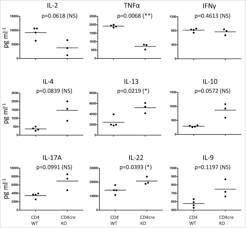
Figure 5.

FURIN-deficient T cells show a Th2- and Th17-type cytokine profile of in the late phase of tumor progression. CD4cre KO and WT mice were treated once with DMBA, then with TPA for 17 weeks (twice a week) on the back skin as in Fig. 1. At 17 weeks, animals were sacrificed and skin draining lymph node cells were cultured for 48 h in the presence of plate-bound anti-CD3 and soluble anti-CD28 antibodies. Cytokine levels were determined from the culture supernatants with ProCartaPlex Mouse Cytokine & Chemokine 26-plex assay and Bio-Plex 200 instrument, or for IFNγ, with ELISA. Statistics: Unpaired two-tailed Student's t-test with Welch correction. CD4+ WT n = 4, CD4cre KO n = 3. Each symbol represents an individual mouse, lines indicate the mean.
