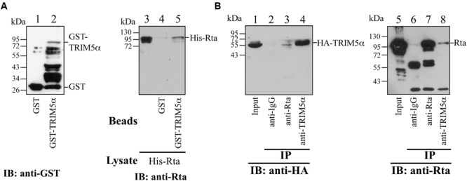
FIGURE 2.

Interaction between Rta and TRIM5α. (A) In a GST-pulldown assay, His-Rta (lanes 3–5) was mixed with GST- (lane 4) and GST-TRIM5α- (lane 5) glutathione-Sepharose beads. Proteins pulled down by the beads were analyzed by immunoblotting (IB) with anti-Rta antibody. Proteins on the glutathione-Sepharose beads (lanes 1 and 2) and Rta in 1% of the lysate (lane 5) were also detected by immunoblotting. (B) Coimmunoprecipitation assay results. Anti-Rta and anti-TRIM5α antibodies were added to the lysate from 293T cells that had been transfected with pLPCX-HA-TRIM5α and pCMV-Rta. Lanes 1 and 5 were loaded with 3% of the cell lysate. Proteins immunoprecipitated (IP) with anti-Rta and anti-TRIM5α antibodies or anti-IgG antibody were detected by immunoblotting (IB), using anti-HA (lanes 1–4) and anti-Rta antibodies (lanes 5–8).
