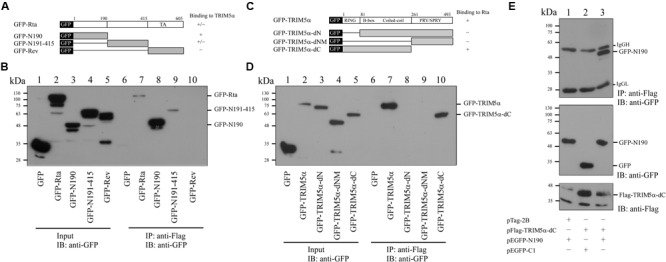
FIGURE 4.

Mapping the interaction domains in Rta and TRIM5α. (A) Plasmids that expressed GFP fused to different segments of Rta were used to delineate the region in Rta that interacts with TRIM5α. (B) 293T cells were cotransfected with pFlag-TRIM5α and plasmids expressing GFP or GFP-Rta fusion proteins (pEGFP-Rta, pEGFP-N190, pEGFP-N191-415, pEGFP-Rev, or pEGFP-C1). Input lanes were loaded with 5% of the lysate (lanes 1–5). Proteins in the lysates were coimmunoprecipitated (IP) with anti-Flag antibody and analyzed by immunoblotting (IB) using anti-GFP antibody (lanes 6–10). (C) Plasmids expressing various GFP-TRIM5α fusion proteins (GFP-TRIM5α, GFP-TRIM5α-dN, GFP-TRIM5α-dNM, GFP-TRIM5α-dC) were generated. (D) 293T cells were cotransfected with plasmids encoding GFP or GFP-TRIM5α fusion proteins (GFP-TRIM5α, GFP-TRIM5α-dN, GFP-TRIM5α-dNM, GFP-TRIM5α-dC). Input lanes were loaded with 5% of the lysate, and GFP-fusion protein expression levels were detected using anti-GFP antibody (lanes 1–5). Proteins in the lysates were coimmunoprecipitated with anti-Flag antibody and analyzed by immunoblotting using anti-GFP antibody (lanes 6–10). (E) 293T cells were cotransfected with pFlag-TRIM5α-dC and plasmids expressing either GFP-N190 or GFP. Input lanes were loaded with 5% of the lysate. Proteins in the lysates were coimmunoprecipitated with anti-Flag antibody and analyzed by immunoblotting using anti-GFP antibody.
