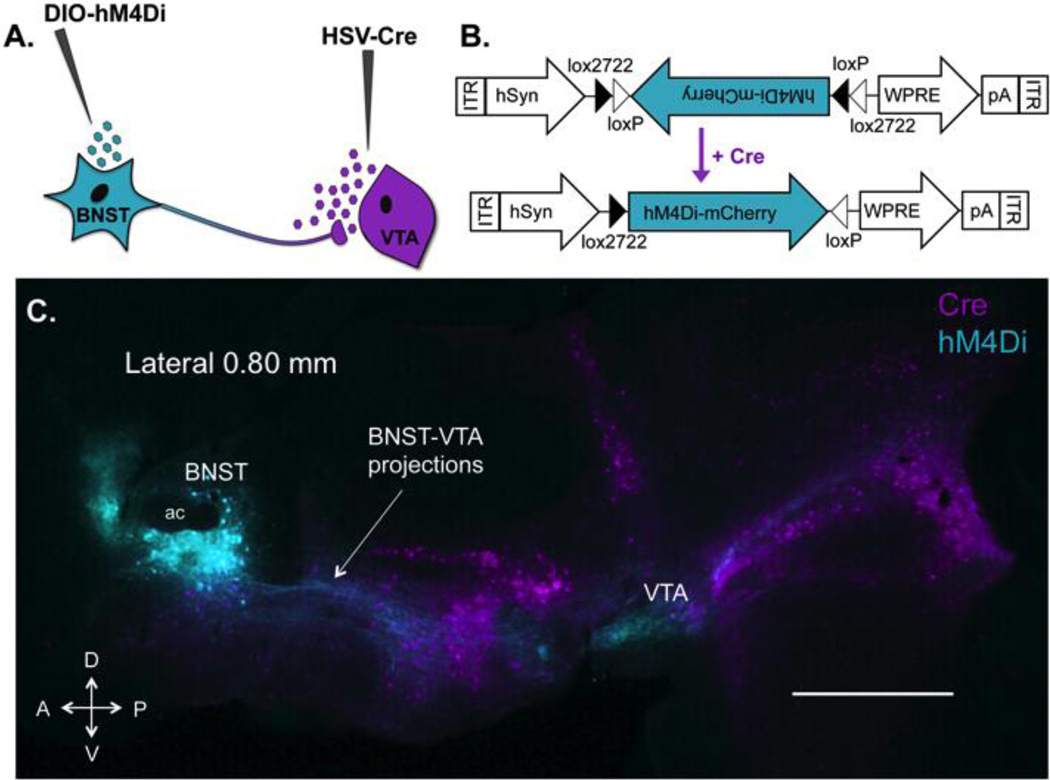
Figure 1. Expression of hM4Di receptors in VTA-projecting BNST neurons using a retrograde intersectional strategy.
(A) A dual-viral approach was used to drive expression of inhibitory designer receptors (hM4Di) in a distinct yet intermixed subpopulation of BNST neurons that project to VTA. A long-term retrograde HSV encoding cre recombinase was delivered into the VTA and a cre-dependent AAV-hM4Di was delivered into the BNST. (B) hM4Di-mCherry AAV vector design employing the double-floxed inverted open reading frame (DIO) strategy. Two pairs of heterotypic, antiparallel loxP-type recombination sites achieve cre-mediated hM4Di inversion and expression under the control of a human synapsin (hSyn) promoter. WPRE, woodchuck hepatitis virus post-transcriptional regulatory element; ITR, inverted terminal repeat, pA, human growth hormone polyadenylation site (C) Sagittal section showing intersection of retrogradely transported HSV-cre (VTA-projecting cre+ cells are pseudocolored magenta) and AAV expressing cre-dependent hM4Di (hM4Di+ BNST-VTA cells are pseudocolored cyan) 8 weeks after vector infusions. Robust expression of hM4Di is visible in soma (in BNST) and fibers (to VTA) of BNST-VTA neurons. HSV-cre transfected neurons projecting to VTA are visible throughout the brain, with the exception of VTA as retrograde HSV does not infect cell bodies at the site of injection. Note the absence of hM4Di in all HSV-Cre transfected cells outside the BNST. This illustrates that hM4Di is localized to VTA projecting neurons within the BNST only. D, dorsal; V, ventral; A, anterior; P, posterior; scale bar, 1 mm.

