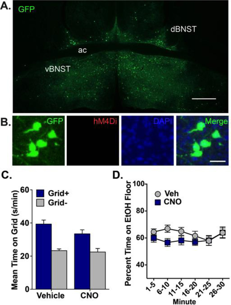
Figure 3. Ethanol CPP expression is not disrupted by CNO in mice expressing GFP in VTA-projecting BNST cells.
(A) Expression of GFP in dBNST and vBNST 8 weeks after infusion of HSV-GFP into VTA and DIO-hM4Di into BNST. GFP+ cells (green) indicate VTA-projecting neurons. No hM4Di expression was visible in BNST. ac, anterior commissure; scale bar, 500 µm. (B) High magnification image from BNST of GFP (green; visualized by IF detection of GFP), hM4Di (absent; visualized by IF detection of mCherry), nuclei (DAPI) and all channels merged. Note that hM4Di is not expressed in the absence of cre; scale bar, 50 µm. (C) Mean (+SEM) time spent on the grid floor (in s/min) during 30-min preference test. CNO did not disrupt ethanol CPP expression in mice expressing GFP in BNST-VTA cells; n=11–12 / conditioning subgroup (Grid+, Grid). (D) There was no significant difference in percent time spent on the ethanol-paired floor between groups when analyzed in 5-min intervals across the 30-min preference test.

