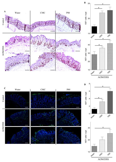
Figure 5. Dietary emulsifiers alter epithelial cell proliferation and apoptosis during colitis-associated cancer development.
(A–B) Epithelial cell proliferation was analyzed by immunohistochemistry using the proliferation marker Ki67 in colonic tissue sections. (A) Representative images of Ki67 staining. Scale bar, 200μm. (B) Ki67+ cells were counted and averaged per crypt. (C–D) Epithelial cell apoptosis was analyzed by terminal deoxynucleotidyl transferase deoxyuridine triphosphate nick-end labeling (TUNEL). (C) Representative confocal images of TUNEL assay: TUNEL, green; DNA, blue. Scale bar, 25μm. (D) TUNEL+ DAPI+ cells were counted and averaged per crypt. Data are the means +/− S.E.M. (n=5–8). Significance was determined using t-test (* indicates p<0.05).

