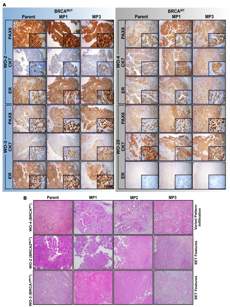
Figure 2. Ovarian cancer epithelial markers and morphologic characteristics in parent tumors are preserved over multiple mouse passages.
(A) BRCAMUT patient-derived-xenograft (PDX) models (WO-2, WO-3) and 2 BRCAWT PDX models (WO-4, WO-20) were evaluated by H&E and immunohistochemistry for epithelial ovarian cancer markers. PAX8 (paired box 8, nuclear stain), CK7 (cytokeratin 7, cytoplasmic stain), and ER (estrogen receptor, nuclear stain) in parent tumor and matched PDXs up to mouse passage 3 (MP1–MP3) are shown. Magnification is ×10 for large panels and ×100 for inserts. (B) Five matched BRCAMUT patient/PDXs and 5 matched BRCAWT patient /PDXs were reviewed for SET (solid, pseudoendometrioid, and translational cell carcinoma-like) morphology in a blinded fashion. WO-4 (BRCAWT), WO-2-1 (BRCA2MUT), and WO-3-1 (BRCA1MUT) representative parent and PDX tumors of MP1, MP2, and MP3 H&E sections shown demonstrating SET criteria in BRCAMUT and not in BRCAWT tumors. Parent WO-2-1 tumor showed micropapillary features, while the MP1–MP3 tumors showed both micropapillary and solid architecture. Magnification is ×10 for all panels.

