
Figure 5. Analysis of circulating tumor cells (CTCs) from 10 patients with metastatic prostate cancer.
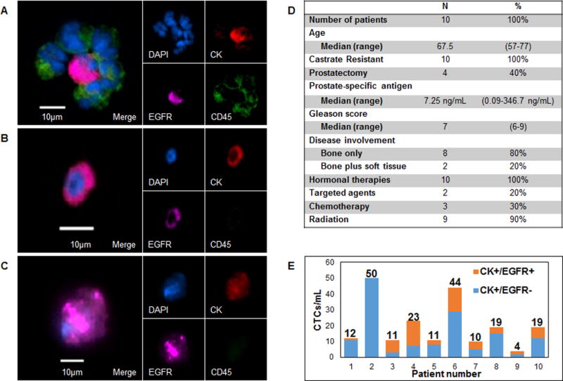
A, Cluster of white blood cells surrounding a CK+/EGFR+ CTC from patient 3. White blood cells stained positive for CD45. B,C, Other representative images of CK+/EGFR+ CTCs from patients 3 and 5 respectively. D, Clinical history of prostate cancer patients analyzed for circulating tumor cells. E, Number of cytokeratin (CK)+/EGFR+ and CK+/EGFR− CTCs isolated per 1mL whole blood. Numbers above the columns indicate the total number of CTCs isolated from patients 1–10.