
Figure 6. Pharmacologic inhibition of HER2 and EGFR is cytotoxic to prostate cancer in vitro and prevents cancer growth in vivo.
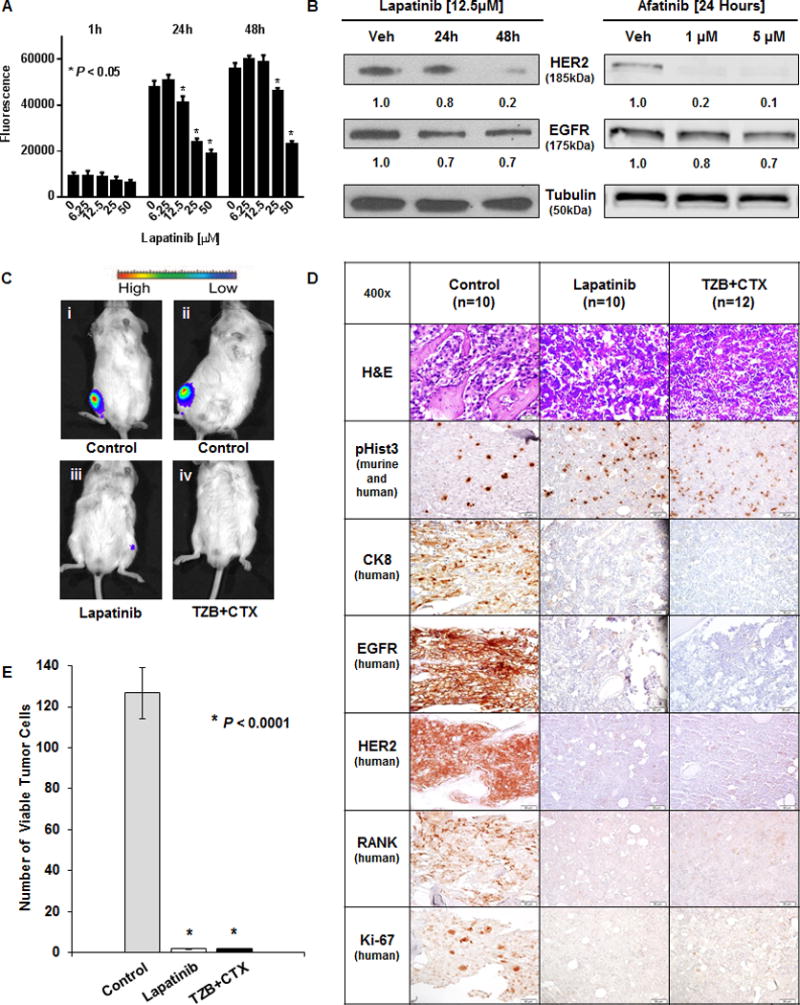
A, C4-2BLuc cell viability measured by Titer Blue following lapatinib treatment for 1, 24, and 48 hours. * denotes P < 0.05 compared with vehicle control. B, (left) Western blot of HER2 and EGFR expression in C4-2B cells following incubation with vehicle or 12.5μM lapatinib at 24 and 48 hours. (right) Western blot of C4-2BLuc cells after incubation with vehicle, 1μM, or 5μM Afatinib for 24 Hours. Tubulin was used as a loading control. (left) Densitometry was performed with ImageJ. (right) Fluorescence was measured using the Odyssey CLx (LI-COR) and quantified using Image Studio v3.1.4 software (LI-COR) (Both) Normalized to Tubulin, values shown under respective lanes. C,D, C4-2BLuc cells were injected into mouse tibiae one week prior to treatments. Untreated mice (Control, n=10), treated with lapatinib (Lapatinib, n=10) or treated with a combination of trastuzumab and cetuximab (TZB+CTX, n=12) biweekly for 6 weeks. C, Bioluminescence of representative mice at the end of the study where i and ii are control while iii and iv received dual inhibitor treatments. D, Serial sections of representative tibia stained with H&E, anti-pHist3, anti-CK8, anti-EGFR, anti-HER2, anti-RANK, and anti-Ki-67. E, Quantitation of viable tumor cells was performed by counting 3 fields of anti-CK 8 positive cells in areas that exhibited Ki-67 positivity in three animals per group. No viable human tumor cells could be found in the lapatinib or in the TZB+CTX groups at 7.5 weeks. * denotes P < 0.0001.