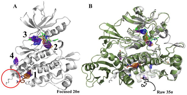
Figure 2.
The first four hotspots from the ABL MixMD maps identify the active and allosteric sites: acetonitrile map (orange mesh), isopropanol map (blue mesh), and pyrimidine map (purple mesh). The active site ligand (PDBid: 3KFA, green) and the allosteric ligand (PDBid: 3K5V, brown) are shown for reference. (A) The four hotspots are shown contoured at 20σ with the spurious sites not shown. The red circle notes the αI′-helix that is referenced in a later section on conformational sampling. (B) MixMD maps of ABL contoured at 35σ with all sites shown. Superimposed on the structure are molecules from the PDB that occupy mapped hotspots on the protein surface. The crystal structure of the full length ABL protein (PDBid: 1OPK, dark green ribbon) was also superimposed to show the SH2 domain interface mapped by the fourth hotspot in MixMD. A different structure (PDBid: 1OPL) has a tyrosine residue (peptide in black) at the packing interface that occupies a small, lower-ranked hotspot at the bottom of the structure.

