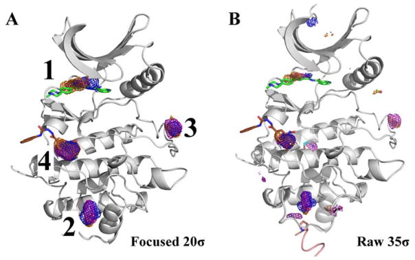
Figure 5.
(A) CHK1 is shown with just the top-four hotspots contoured at 20σ. Maps are shown in mesh: acetonitrile (orange), isopropanol (blue), and pyrimidine (purple). The first and the fourth hotspots map the active (PDBid: 1ZYS, green) and allosteric site (PDBid: 3JVS, brown). The second and the third-ranked hotspots map sub-sites in the binding groove for the peptide substrate. (B) Maps contoured at 35σ show lower-ranked hotspots overlap with different CHK1 structures showing protein-packing interfaces (contact in structure 2YDK is shown in pink) and small crystal additives (structures 4FTO in black, 2YM8 in cyan, and 2XF0 in dark green).

