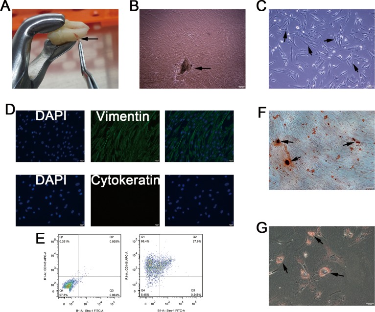
Fig 1. Isolation and characterization of human PDLSCs.
(A) We isolated PDLCs from PDL tissues (arrow), which are attached to the surface of the roots. (B) Cell clusters, derived from tissue blocks (arrow) showed radiating or whirlpool-like morphology. (C) Isolated CD146+ PDLCs were small, round, fusiform, and triangular (arrow). The cells had abundant cytoplasm and large nuclei, and several binucleate cells were observed (arrow). (D) Immunofluorescence staining showed that isolated CD146+ PDLCs stained positive for vimentin, and negative for cytokeratin. (E) Flow cytometry analysis showed that the percentage of cells expressing the stem cell surface markers STRO-1 and CD146 was 27.9%, after CD146 immunomagnetic isolation. (F) Alizarin Red staining showed the formation of some mineralized nodules in cultured PDLSCs after osteogenic induction for 3 weeks. (G) PDLSCs formed Oil Red O-positive lipid droplets after 3 weeks of induction in the adipogenic medium.

