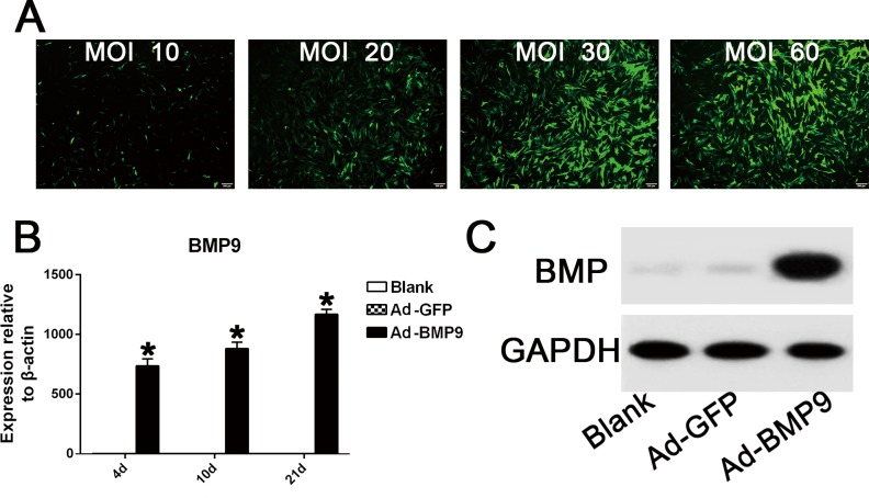
Fig 2. Helper GFP infection efficiency and overexpression of BMP9 in hPDLSCs following BMP9 transfection.
(A) The fluorescence area of GFP-positive cells increased gradually when the MOI value ranged from 10 to 30, while the GFP infective efficiency remained similar between the 30 MOI group and the 60 MOI group. Magnification, 100×. (B) qRT-PCR results showed that the BMP9 transfection group had 700–1200 fold higher gene overexpression of BMP9 compared to the control GFP group. “*”, p<0.01 (vs. control groups). (C) Western blot assay suggested that in the Ad-BMP9 transfection group, BMP9 protein expression was significantly promoted until day 21.

