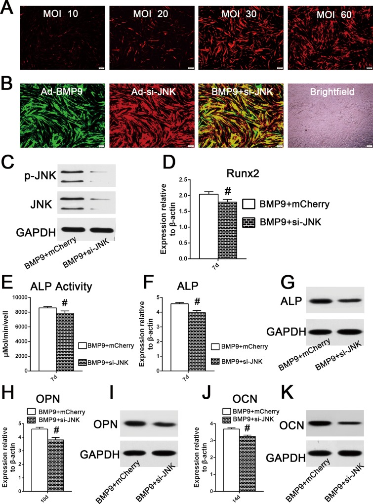
Fig 5. JNKs knockdown decreased BMP9-stimulated osteogenic differentiation of hPDLSCs.
(A) The helper mCherry infective efficiency was determined by the observation of the area of fluorescence after hPDLSCs were transfected with different adenoviral concentrations (10, 20, 30, or 60 MOI) for 2 days. (B) When hPDLSCs were co-infected with Ad-GFP (MOI 30–60) and Ad-mCherry (MOI 30–60), the co-infective efficiency was approximately 80%, according to observation of the area of fluorescence 72h post co-infection. (C) Western blot analysis showed that si-JNK significantly decreased the phosphorylation of JNK1/2 in hPDLSCs stimulated by BMP9, after 72h of co-infection. (D) Effect of JNK1/2 gene silencing on the mRNA expression of Runx2 in hPDLSCs stimulated by BMP9, determined by qRT-PCR analysis at day 7. ‘‘#”, p<0.01 (vs. BMP9 group). (E) and (F) and (G) Effect of JNK1/2 knock down on the gene and mRNA expression of ALP in hPDLSCs stimulated by BMP9 by a quantitative ALP activity kit, qRT-PCR assay, and western blotting. ‘‘#”, p<0.01 (vs. BMP9 group). (H) and (I) Effect of JNK1/2 gene silence on the mRNA and protein expression of OPN in hPDLSCs stimulated by BMP9, was determined by qRT-PCR and western analysis at day 10. ‘‘#”, p<0.01 (vs. BMP9 group). (J) and (K) Effect of JNK1/2 gene silencing on the mRNA and protein expression of OCN in hPDLSCs stimulated by BMP9, was determined by qRT-PCR and western analysis at day 14. ‘‘#”, p<0.01 (vs. BMP9 group).

