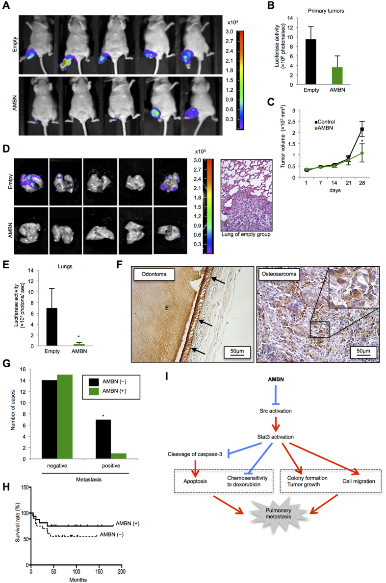
Figure 4. AMBN inhibits tumor growth and pulmonary metastases in vivo, and AMBN expression is correlated with pulmonary metastasis and prognosis in human osteosarcoma cases.
(A) Photons from luciferase in primary tumors of empty and AMBN groups after 28 days were captured. (B) Quantification of photons from luciferase in the primary tumors is shown (N = 5). (C) The volume of primary tumors at indicated days is shown (N = 5). (D) Photons from luciferase in the lungs after 28 days were captured (left panels) and representative metastatic focus of lung in empty group is shown (right panel). Original magnification of the right panel: ×200. (E) Quantification of photons from luciferase in the lungs is shown (N = 5). (F) AMBN expression in 37 osteosarcoma cases was evaluated by immunohistochemical analysis. Specificity of anti-AMBN antibody was confirmed by using odontoma. AMBN protein was stained in ameloblasts and enamel. Arrow indicates ameloblasts and “E” indicates enamel (left panel). Representative tissue of AMBN staining in osteosarcoma cases is shown. AMBN was expressed in cytoplasm of osteosarcoma cells (right panel). (G) The correlation between AMBN expression and pulmonary metastasis is shown (N = 37, chi-squared test). *P < 0.05. (H) The correlation between AMBN expression and survival rate for 37 patients with osteosarcoma was analyzed and shown as a Kaplan-Meier graph (N = 37, log-rank test). (I) In this model, AMBN inhibits Src activation (dephosphorylation at Y416) followed by Stat3 inactivation (dephosphorylation at Y705). Continuous inactivation of the Src-Stat3 pathway induces caspase-3 mediated apoptosis and chemosensitivity to doxorubicin, while suppresses colony formation, tumor growth and cell migration. These activities lead to the reduction of pulmonary metastasis. Mean ± SEM (B,C,E); *P < 0.05.

