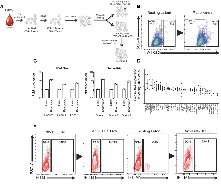
Figure 1. Generation of latently infected CD4+ T cells.
(A) CD4+ T cells were negatively selected from PBMCs and conditioned for 3 days with CCL19. CCL19-treated cells were infected with HIV-1NL4-3 for 6 days, and resting CD4+ T cells (CD25–CD69–HLA-DR–) were negatively selected by magnetic cell sorting. Resting CD4+ T cells were reactivated with anti-CD3/CD28 for 3 days. (B) Representative intracellular staining of HIV-1 Gag (KC57) (n = 3). (C) Quantification of HIV-1 gag and mRNA transcripts by real-time qPCR. (D) Fold expression of selected antiviral genes in resting latent relative to reactivated cells. Data are plotted as mean ± SEM where n = 3. (E) Representative extracellular staining of IFITM1 (n = 3).

